DISCOVERY ACROSS DISCIPLINES IN NEUROSCIENCE AND PHYSICS:
FINNISH INNOVATION IN BRAIN IMAGING
Riitta Hari: ‘We shared a common goal: understanding how the human brain works’
When neuroscientist Riitta Hari started working at the Low Temperature Laboratory of the Helsinki University of Technology, methods to measure brain activity were still very limited. Over the next decade, brain research and the instrument development stimulated and supported each other for continuous progress.
In early 1981, Riitta Hari, who had recently earned her Doctor of Medicine degree, received a surprise job offer from Professor Olli Lounasmaa, who founded the Low Temperature Laboratory in 1965. The laboratory was a world leader in ultra-low temperature research and renowned for record-low temperatures, reaching nearly absolute zero.
Lounasmaa had recently become interested in human brain function, which he considered a scientific problem comparable to the Big Bang. He felt that the team of physicists would benefit from a neuroscientist, and so he invited Riitta Hari to work at the Low Temperature Laboratory. Hari agreed to leave the hospital as soon as she had completed her Specialist Degree in Clinical Neurophysiology and secured her own funding. ‘I didn't want to be bossed about by anyone,’ she says.
Company founded by researchers becomes market leader
In September 1982, Hari started to work at the Low Temperature Laboratory and head the small research group studying human brain function using magnetoencephalography,or MEG.
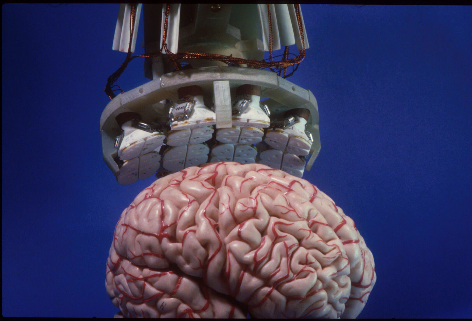
MEG shows when and where groups of brain cells are active. The method measures the magnetic fields produced by neuronal currents with a precision of milliseconds. The equipment uses highly sensitive SQUID sensors, which are kept superconducting by immersing them in liquid helium at a temperature of –269°C.
Riitta Hari had already used single-channel MEG equipment to measure auditory responses during her dissertation. However, the equipment couldn’t provide information from the whole brain at once. This prevented the study of more complex phenomena and, for example, spontaneous brain activity.
The Low Temperature Laboratory invested heavily in developing multi-channel MEG equipment. A 4-channel device was completed in 1984, a 7-channel device in 1986 and a 24-channel one in 1989.
‘At the Low Temperature Lab, instrument development and neuroscientific research went hand in hand, which was of vital importance. When we gained access to better equipment, we carried out measurements around the clock and used the new results to justify why the equipment should be developed further,’ Hari says.
In 1989, Lounasmaa, Hari and three of their colleagues founded a company named after the MEG sensors – the Finnish word mustekala translates to squid.
Three years later, Mustekala, which later became Neuromag Oy, built the world's first whole-head MEG device with 122 channels. In three decades, the company has grown, changed names and ownership, and become the world leader in its field.
These days, MEG is widely used in both basic research and medical diagnostics, such as locating epileptic foci before surgery.
Bringing neuroscience and physics under the same roof raised a few eyebrows in the 80s, but it was worth it. ‘We shared a common goal: understanding how the human brain works. Such a convergence-research approach went far beyond multi- and interdisciplinary approaches by integrating diverse perspectives, insights and knowledge on a common problem,’ Riitta Hari says.
Text by Minna Hölttä
Sensors using quantum optics may revolutionise the way brain signals are measured
Lauri Parkkonen and his team are building a novel high-resolution MEG device whose sensors are much closer to the brain than in conventional MEG. It opens up new possibilities for diagnosing brain injury, cognitive decline, dementia, and other conditions, and strengthens Finland's position as a forerunner in the field.
In the early 2010s, Professor Lauri Parkkonen started to think about solving the fundamental problem of MEG, a device used to measure brain activity.
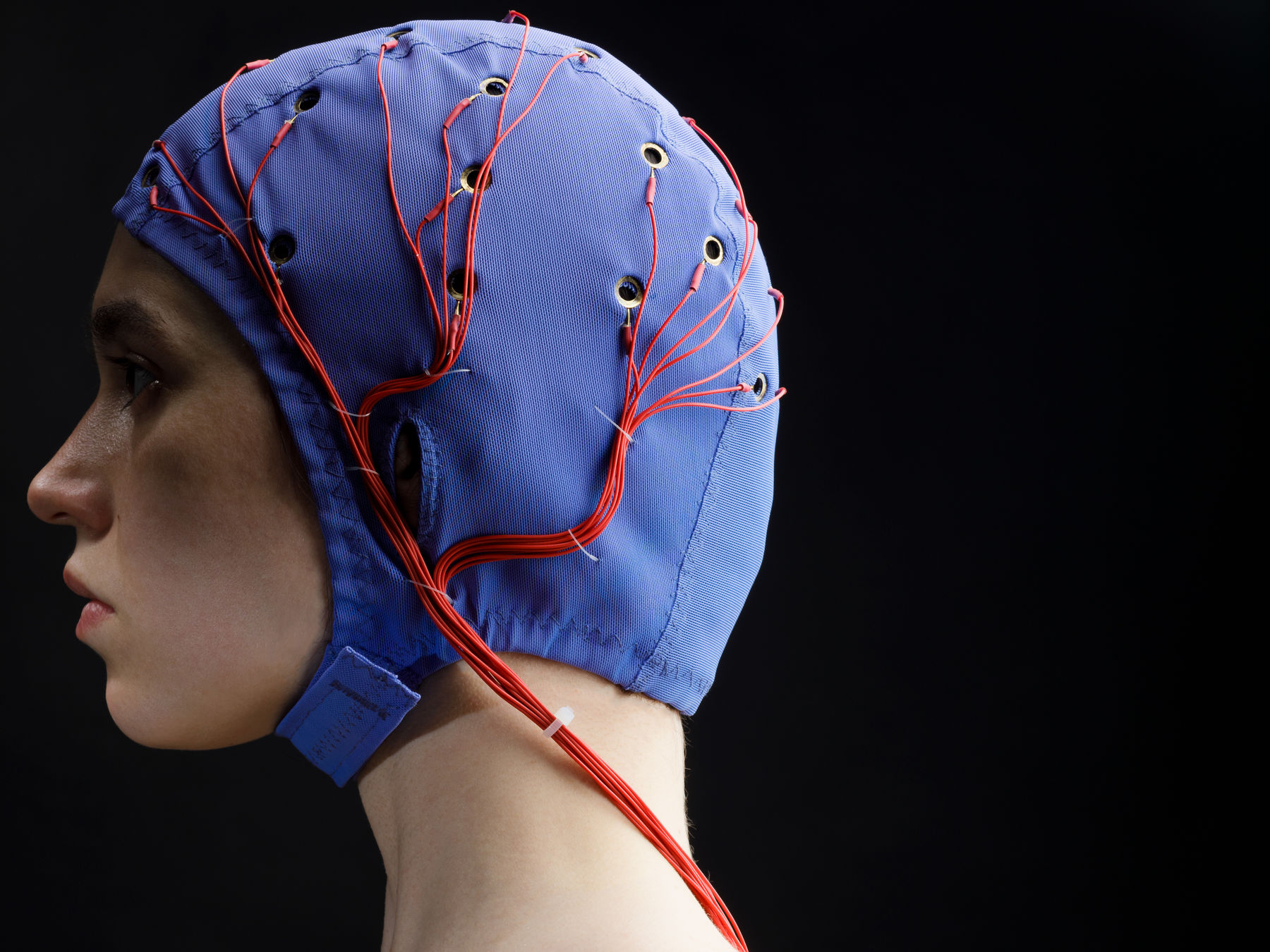
The SQUID sensors used in MEG or magnetoencephalography scanners need to be extremely cold to function, just a few degrees above absolute zero. That’s much colder than a human head, so the sensors have to be properly insulated, which adds to the distance between the sensors and the head. In addition, with such sensors, the sensor helmet cannot be adapted to the head size; the smaller the head, the further away from the surface of the scalp the sensors are – and the trickier it is to perform accurate measurements.
‘This means that with babies and children, we can only measure one brain lobe at a time,’ Parkkonen says.
Parkkonen learned about new sensors based on quantum optics and realised he had found a potential solution. The individually packaged sensors function at room temperature and can be placed within a couple of millimetres of the scalp, measuring stronger signals and making it easier for the researchers to distinguish activity in brain areas that are close together.
The European Research Council (ERC) granted funding for the project, enabling the research group to start building the device. Parkkonen and his team purchased the sensor components from the United States but made everything else themselves, from the sensor helmet to the electronics and software. They sawed and 3D printed parts both at Aalto workshops and in Parkkonen's garage.
‘Carpentry has turned out to be a useful hobby,’ Parkkonen says, laughing.
Getting closer to hospital use and patient measurements
The work has progressed quickly, and the results are promising: the accuracy of the measurements made using optical sensors is nearly the same as with sensors inside the skull.
The next step is to take the device from the lab in Otaniemi to the BioMag laboratory at Meilahti Hospital. If measurements done on healthy test subjects go well, the group will start planning the first tests on patients.
MEG imaging isn’t used only in basic research but also to locate epileptogenic zones and map important areas of the cerebral cortex before brain surgery. The researchers believe that MEG could one day be used to help diagnose early-stage Alzheimer's and recognize functional changes in the brains of patients with brain injury, for example.
Parkkonen is also involved in the macQsimal project, part of the European Union’s one-billion euro Quantum Flagship initiative, which strives to develop the optical sensors further. The project's participants also include the leading MEG device manufacturer Megin Oy, founded in Otaniemi, in whose early stages Parkkonen himself participated actively.
Parkkonen believes that MEG devices based on optical sensors may turn out to be slightly less expensive than traditional equipment – and easier to place, too, since they need a smaller space that’s protected from magnetic fields. ‘This would make MEG accessible for more researchers and hospitals.’
Text by Minna Hölttä